By Rory Dulock | Staff Writer
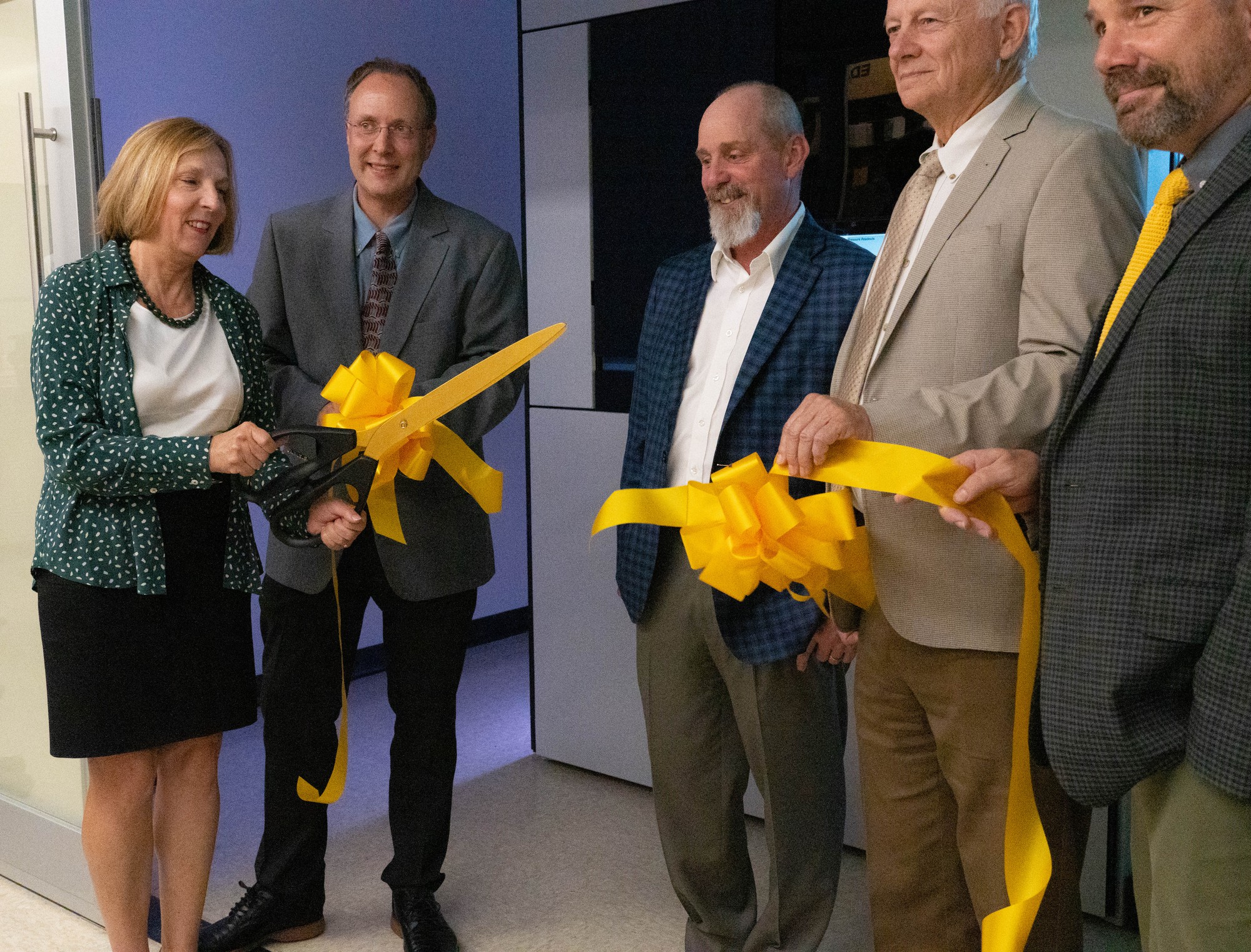
After three years and $8 million, the Baylor Center for Microscopy and Imaging presented its transmission electron microscope Tuesday at the Baylor Sciences Building.
The Baylor Center for Microscopy and Imaging held a ribbon cutting and demonstration for the recently acquired $8 million microscope. Dr. Bernd Zechmann, director of the Center for Microscopy and Imaging, said the transmission electron microscope has multiple uses and benefits.
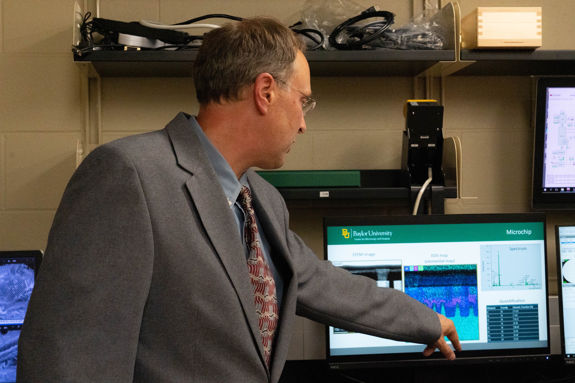
“The transmission electron microscope uses electrons that travel through the column and then shoot through your samples, and then on the bottom, you see different contrasts,” Zechmann said. “The electrons that go through are bright, and the ones that don’t go through will be black. So this microscope can give you a quick black and white picture of your sample, and if you have a live sample, you can see matter from germs or membranes or endoplasmic reticulum or ribosomes.”
Zechmann said the transmission electron microscope is one of only two of its kind in the entire world.
“When I started my job as the director of the Center for Microscopy and Imaging in 2014, I noticed that the, back then, 18-year-old transmission electron microscope was not suited for tier one research,” Zechmann said. “I spent the first two years meeting with faculty, students, companies and also the faculty administration … to find the timeline to replace the microscope and also funding options.”
The process to order, receive and build the transmission electron microscope took a couple of years, Zechmann said.
“We ordered it in 2020, and then they had to assemble it … and then they did all of the testing, and then they shipped it over, and we received it in July 2022,” Zechmann said. “From July 2022, it took them until May 2023 to assemble it, and then we signed off on it in May 2023.”
The acquisition of the transmission electron microscope would not have been possible without the collaborative effort between different departments at Baylor, Zechmann said.
“In the years between 2016 and 2020, there was a truly remarkable collaborative effort of 11 major and 14 minor research groups that represented over 150 potential users to replace the old transmission electron microscope,” Zechmann said. “This microscope is truly a collaborative effort of all these faculty that have worked together to bring it to Baylor University.”
Zechmann said the “future will be very bright with this microscope,” as plans are already being made for its usage.
“We’ll train all of the users on the new microscope and transition them over in the summer,” Zechmann said. “By fall, we’ll hopefully get advanced training on tomography. We’re in the process of hiring an electron microscopy specialist … so [students] will be able to use all the capabilities of the microscope.”
McGregor doctoral candidate Kayla Haberman was the first student trained to use the transmission electron microscope. She said a person has to be mindful of the electron beam that the microscope has.
“Not only are you aware of the electron beam, you get to move this beam within your sample to view different parts of the cell,” Haberman said. “You want to find where you’re at in your grid, and you want to find a cell, and then you want to increase your magnification and zoom in on it.”
Haberman said that to operate the microscope, students are required to take the transmission electron microscope class, which is only offered to graduate students.
“Dr. Zechmann will take an entire semester to teach you how to prepare your sample, how to section your samples,” Haberman said. “We treat our sample with different chemicals that will allow for the contrasts to be seen, so it will bind to different membranes to allow those membranes to be seen. We are able to visualize what we collected, what your sample was — either particles and metals and different atoms — or you can look at different biological structures.”
The transmission electron microscope is able to show the reality of what cells really look like as opposed to how they are portrayed in textbooks, Haberman said.
“It’s really cool to see the actual structure of cells and how it’s a little different from what we get taught in school,” Haberman said. “It’s just super cool to come full circle from treating the cells to observing them on the TEM [transmission electron microscope] and seeing these structures are real.”